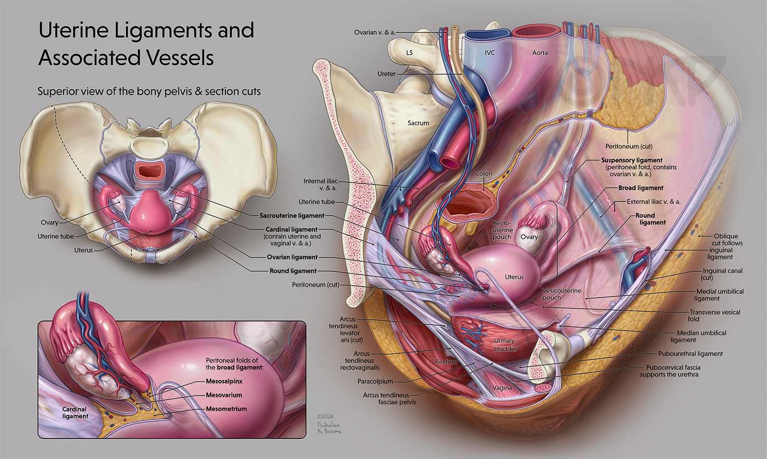
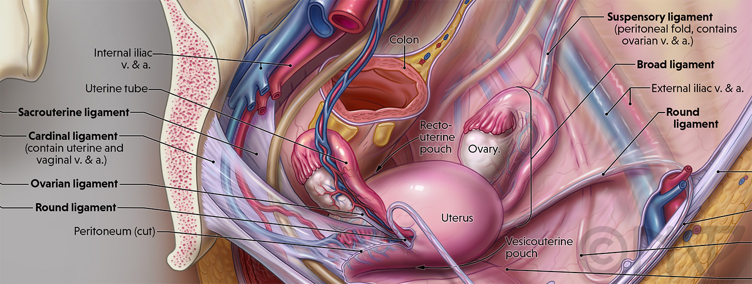
This anatomical plate illustration highlightsall the uterine ligaments (bold labels), as well as vasculature they are associated with. It is meant to help medical students study this difficult area of anatomy. The first callout shows a superior view of the uterine ligaments within the pelvis and the location of the cross-section cuts in the main image. The main image is a three quarters view of the ligaments within the pelvis and the vasculature associated with them.The angle of the main image was chosen to portray all the ligaments with and without the peritoneum covering them. This image allows the viewer to appreciate a more three-dimensional view of the ligaments. The second callout set at the same angle as the main imageclarifies the peritoneal folds of the broad ligament.