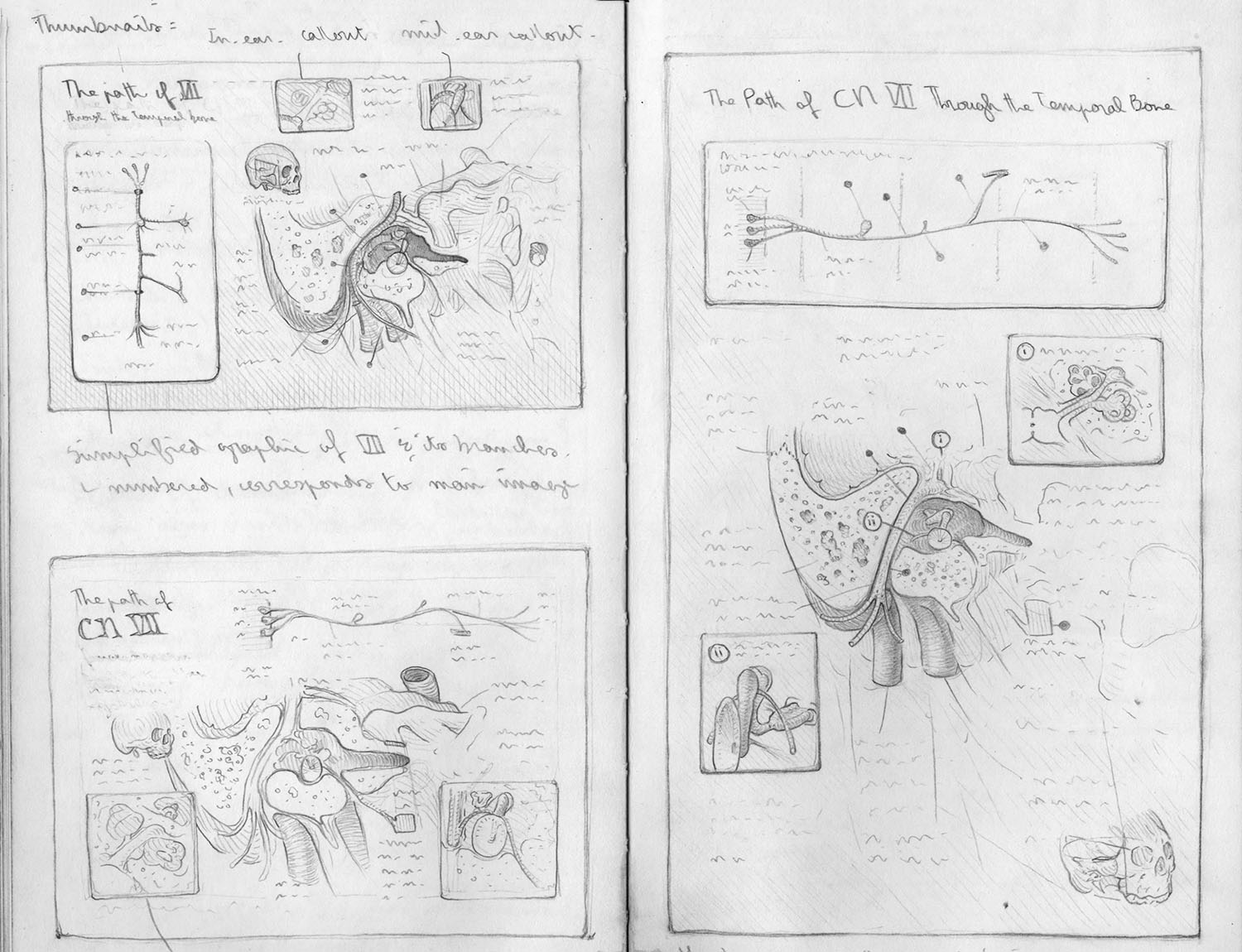
The Path of the Facial Nerve Through the Temporal Bone
Click any image to enlarge
Full poster
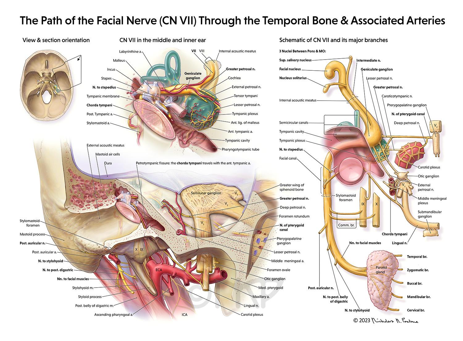
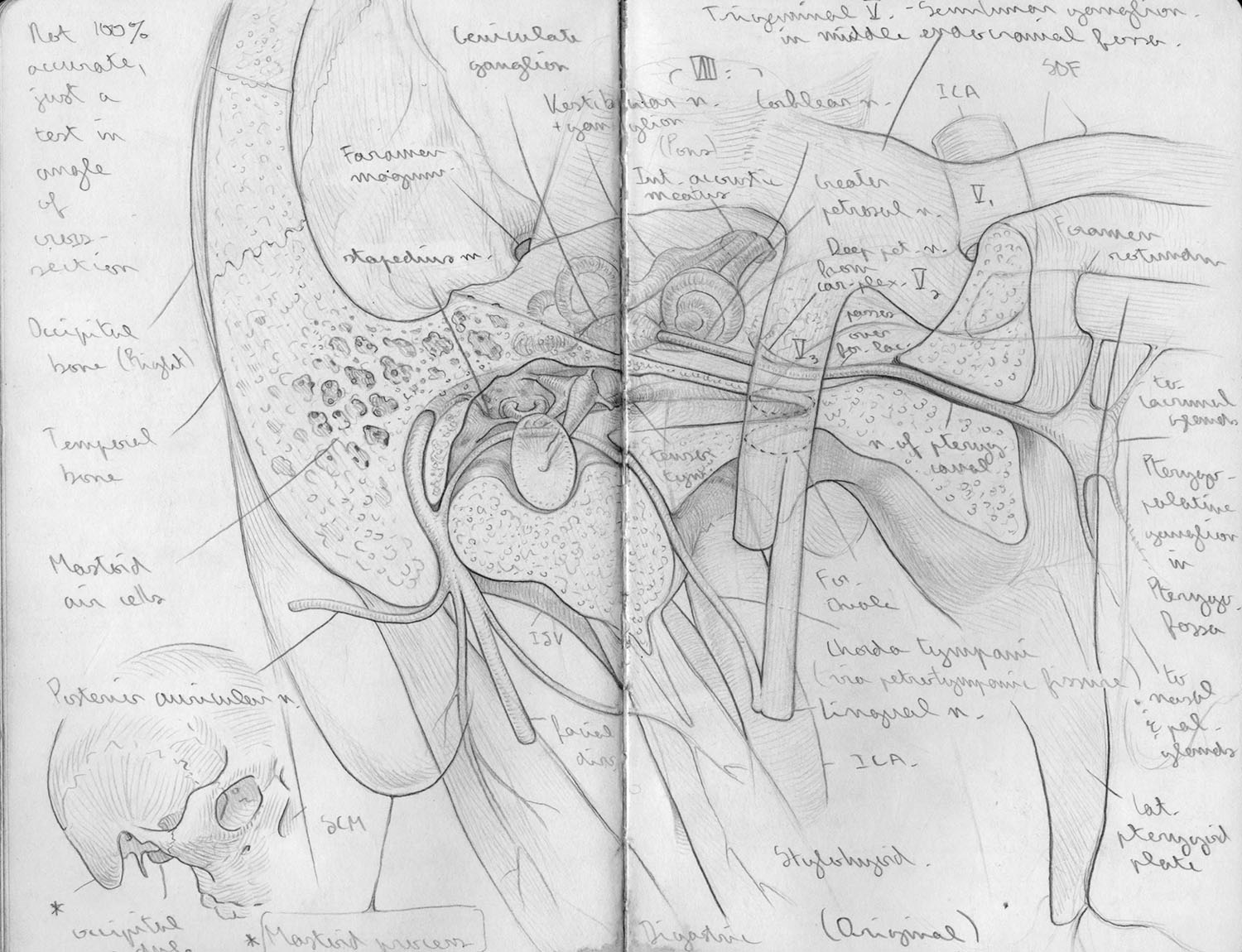
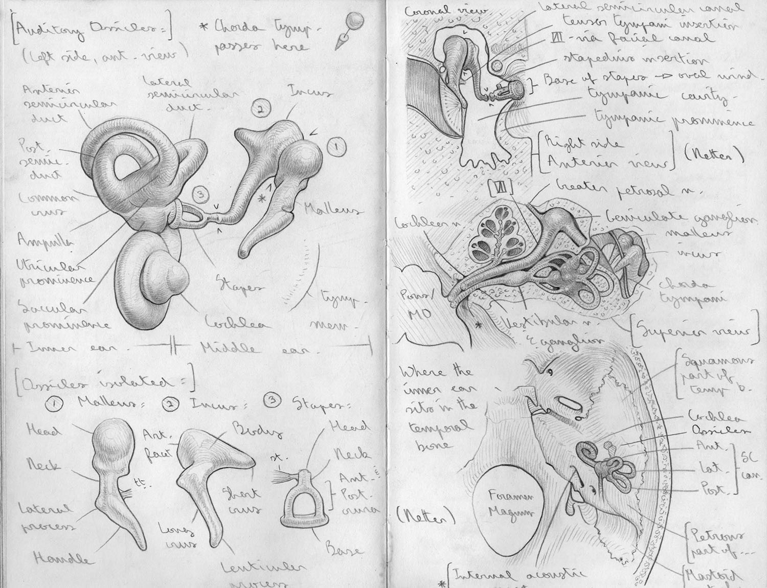
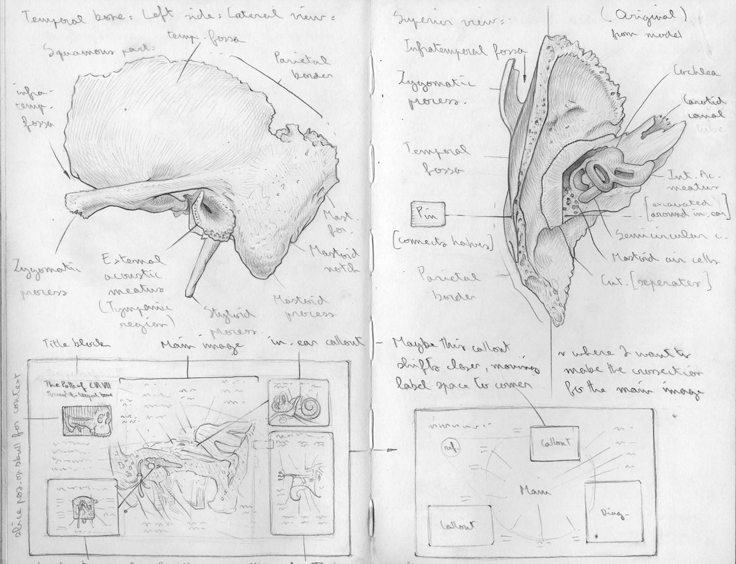
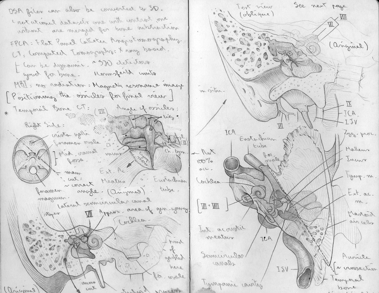
This anatomical plate was designed for a first year medical student, it illustrates the path that the facial nerve (CNVII) takes through the temporal bone. The superior, axial view of the calvarium describes the angle at which the bone has been cut to best view the anatomy.
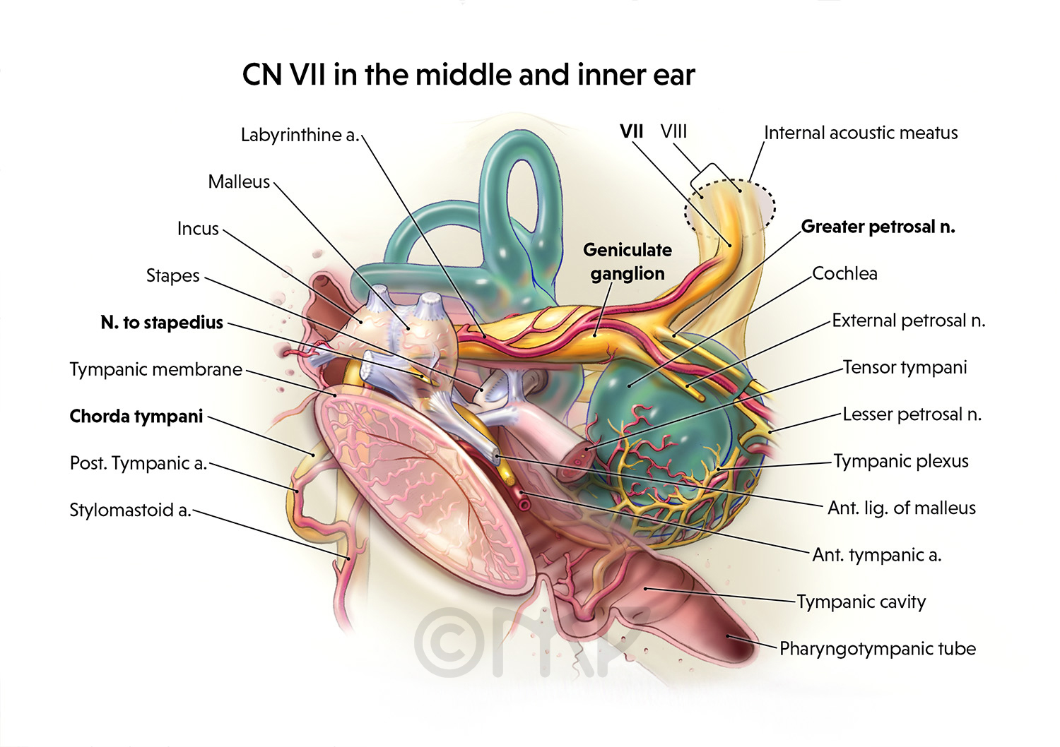
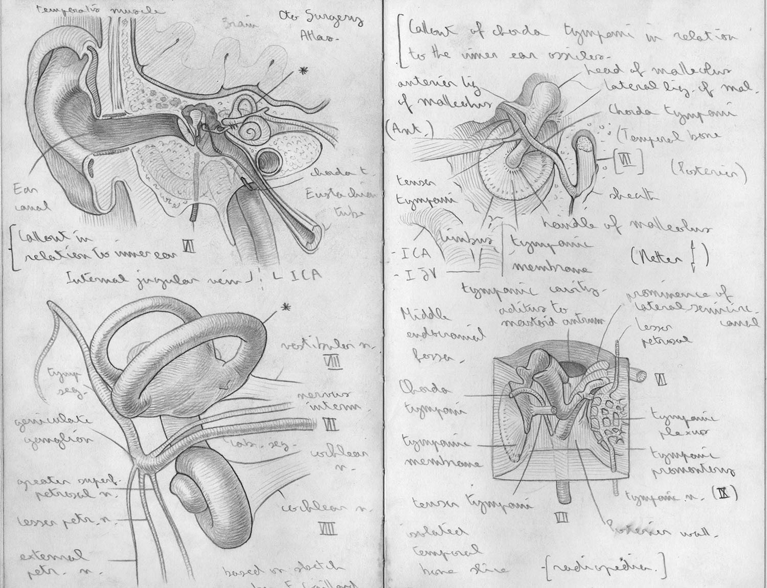
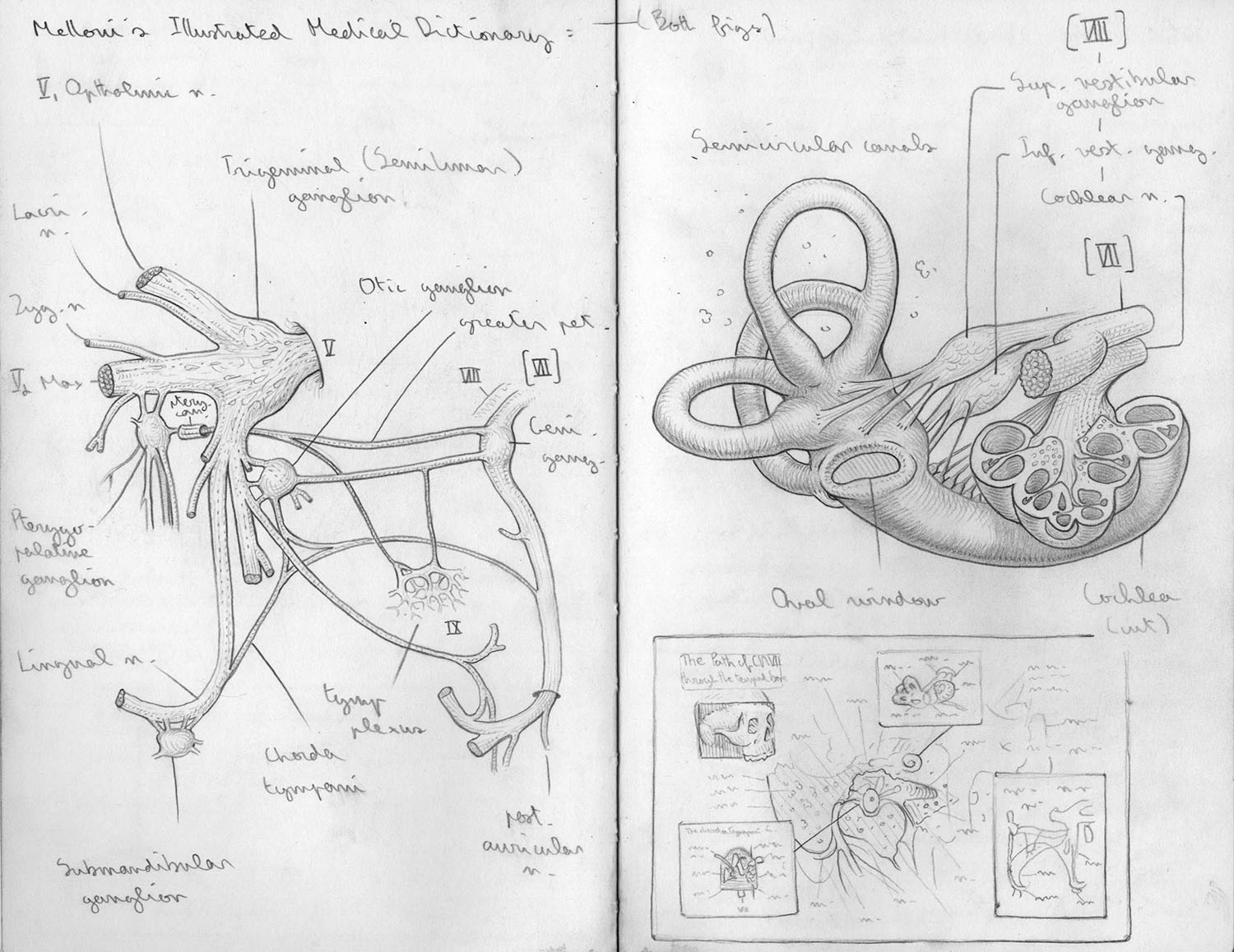
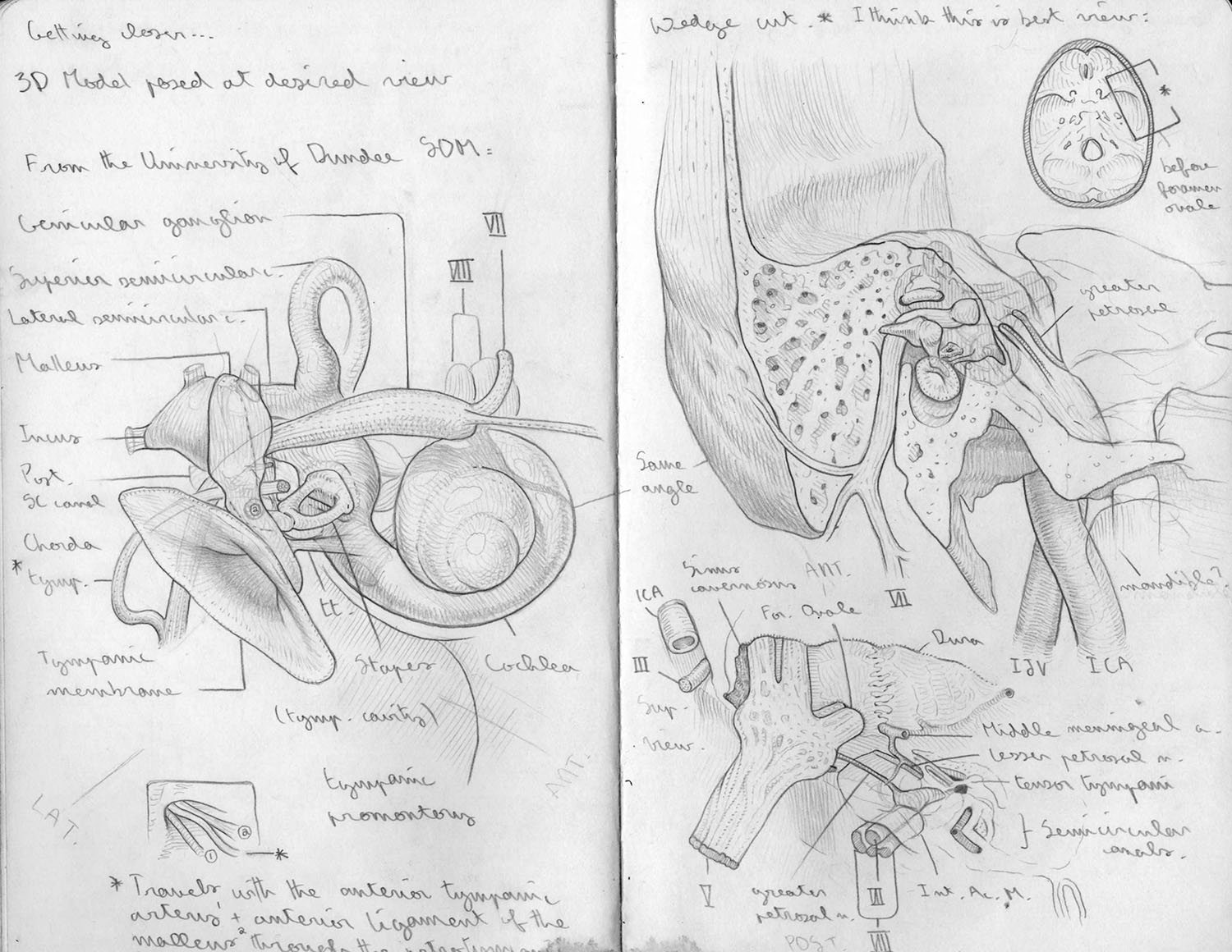
The first image highlights CN VII’s close association with CN VIII as it travels around the inner ear forming the geniculate ganglion, then diverts to travel through the middle ear to the facial canal, sending off branches as it goes that interact with the tympanic plexus and ossicles.
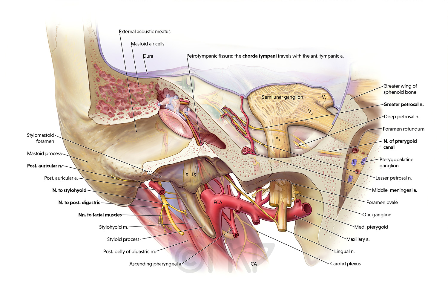
The main image illustrates these and other major branches of CN VII, as well as the bony landmarks, foramen, arteries, and other CNs it networks with, since these subjects are often learned simultaneously. It also illustrates the muscles it innervates before reaching the parotid gland.